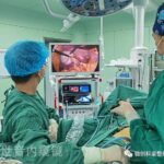
Laparoscopy: Dimensional Surgery Supine Right Pelvic Lymphadenectomy Plus Bilateral Retroperitoneal Lymphadenectomy
Fully Exposing The Following Parts During Operation
- Abdominal aorta and its branches superior mesenteric artery, left and right renal artery, inferior mesenteric artery, left and right reproductive artery, lumbar artery, bifurcation of abdominal aorta, right common iliac artery, right internal iliac artery, umbilical artery, right external iliac artery;
- Inferior vena cava and its branches left renal vein, lumbar vein, right common iliac vein, right internal iliac vein and right external iliac vein;
- Obturator nerve;
- Right ureter;
- Right spermatic cord;
- Others include right obturator vessel, inferior mesenteric vein, left reproductive vein, left ureter, duodenum, lymphatic vessel in front of left renal vein, diaphragm foot, psoas major muscle, iliopsoas muscle, pelvic fascia, etc.
Operation Steps: According To The Procedure Of Dimensional Surgery
1. The peritoneum was cut along the mesenteric root to establish the digestive and urogenital plane (in fact, the embryonic retroperitoneal plane), and the peritoneum margin was lifted under the upper abdominal wall with a band of hemolok to remove the bowel from the urogenital cavity and the large vascular cavity.
2. The genital space is located in front of the urinary space, so the spermatic cord fascial space was removed from the ureteral fascial space.
3. The ureteral fascial space was established and the ureter was retracted to expose the common iliac vessels and the right pelvic wall.
4. Right pelvic lymphadenectomy was performed first. The lymphoid layer includes the perivascular and nerve planes, musculoskeletal fascia planes.
The idea of level surgery is to establish the peripheral plane of blood vessels and nerves, that is, to free the blood vessels and nerves that need to be protected from the lymphoid adipose tissue in advance.
In order to avoid bleeding polluting the operation field, from low altitude to high altitude, the order is anterior and lateral planes of internal iliac artery and vein, peripheral plane of obturator nerve, peripheral plane of external iliac artery, peripheral plane of external iliac vein, peripheral plane of common iliac artery and vein, and plane of iliopsoas muscle and obturator muscle.
After the establishment of these planes, the pelvic lymphoid adipose tissue was paged, and the leaf ridges were cut and cleaned step by step.
5. Then the lymphoid adipose tissue from the right common iliac artery to the starting point of the inferior mesenteric artery was dissected. The following planes were established in turn: the abdominal aortic sheath was cut longitudinally to establish the plane around the abdominal aorta;
The inferior vena cava sheath was dissected longitudinally to establish the plane around the inferior vena cava;
The lamellar lymphoid adipose tissue between the main abdominal cavity and the inferior lumen was cut and excised;
The medial plane of the right psoas major muscle was established, and the lateral lymphoid adipose tissue of the paged inferior cavity was cut and excised;
The medial plane of the left psoas major muscle was established, and the paged ventral main lateral lymphoid adipose tissue was cut and excised.
In the above steps, the monitor is on the foot side, and then move the monitor to the head side.
6. Expand the retroperitoneal plane of the embryo, supplement the peritoneum margin lifting to isolate the intestinal tube in front of the embryo peritoneum, and expose the cephalic inferior vena cava, left renal vein, abdominal aorta, left and right reproductive vessels and ureters.
7. Continue to follow the following ideas:
- Establish the plane around the inferior cavity, ligate and cut off the right reproductive vessel at the root;
- Expand the plane around the inferior cavity to the plane around the left renal vein;
- The abdominal aortic sheath was dissected longitudinally to establish the periaortic plane;
- The medial plane of the left reproductive vessel was extended to the left renal vein;
- The enlarged lymphoid adipose tissue on the lateral side of the abdominal aorta was excised from the plane of the diaphragm foot and psoas major muscle;
- The lymphoid adipose tissue between the main abdominal cavity and the inferior lumen was excised from the vertebral body plane;
- The lymphoid adipose tissue on the right side of the inferior vena cava was excised from the vertebral body plane and the medial plane of the psoas major muscle.
The idea of the above steps is to first establish the left and right planes of lymphoid adipose tissue to page it, and then cut the paged lymphoid adipose tissue in the musculoskeletal plane.
Summary
The peripheral layers of lymphoid adipose tissue include: Retroperitoneal plane (digestive and urogenital plane), periarterial plane, perivenous plane, perineural plane, musculoskeletal fascia plane. The idea of level surgical lymph node dissection is not to strip the lymphoid adipose tissue from the surface of blood vessels and nerves, but on the contrary, first free the blood vessels and nerves that need to be protected from a pile of lymphoid adipose tissue through the surrounding plane of each pipeline itself, and then page the lymphoid adipose tissue and cut it from the musculoskeletal fascia plane.
Pelvic lymph node dissection procedure
Step #1
After successful anesthesia, remove the abdominal median or paramedian incision, the incision reaches 3-5 cm above the umbilical cord, cut the skin and subcutaneous successively, separate the rectus abdominis muscle, and expose the peritoneum.
Lift the edge of the anterior sheath of the right rectus abdominis muscle, and gently separate the right extraperitoneal space with the palms of the four fingers of both hands close to the peritoneum, so as not to damage the inferior arteries and veins of the abdominal wall. At the level of the anterior superior iliac spine, the round ligament was found at the internal opening of the inguinal canal, and the clamp was broken. The No. 7 suture was ligated, and the suture was retained at the proximal end.
Step #2
The psoas major muscle, iliac vessels and ureter of the retroperitoneum were exposed by pulling the suture cut at the edge of the retroperitoneum on the right side, and the surrounding tissues were dissociated along the ureteral route. Pull the hook to open the upper peritoneum and external abdominal wall to expose the operation field. The adipose connective tissue of psoas major muscle was separated to expose the genitofemoral nerve.
The lymphoid adipose tissue in front of and around the right iliac and external iliac arteries was removed passively and sharply from top to bottom. Continue to free the lymphoid adipose tissue in front of the external iliac vein and pull outward to expose the internal iliac artery and separate the lymphoid adipose tissue between them. The deep inguinal lymph nodes were separated at the lowest end of the iliac vessels, and the distal end reached the intersection of circumflex iliac veins.
Step #3
Pull the bladder to the inside, pull the hook to open the external iliac vein, enter the obturator fossa, and separate the obturator nerve. Above the obturator nerve, the obturator fossa lymphoid adipose tissue was separated from bottom to top. So far, pelvic iliac vessels, inguinal and obturator lymph nodes have been cleared. With the same method, the left pelvic lymph nodes were cleaned and the internal iliac artery was ligated to reduce the bleeding during hysterectomy. The operation should pay special attention to the details, and each link is particularly critical.
Thanks for reading, we hope you have enjoyed the content.
If you are looking for the endoscopy equipment, feel free to check our product page or contact us at our contact page.


Leave a Reply
Want to join the discussion?Feel free to contribute!