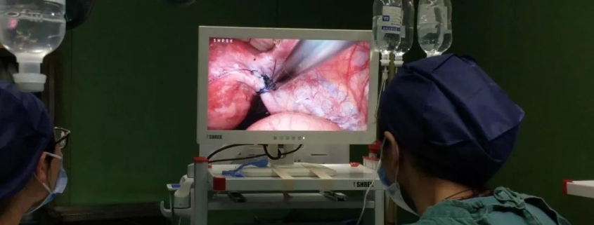
Laparoscopy: Conservative Surgery for Tubal Pregnancy
Indications
1. Have fertility requirements, especially those whose contralateral fallopian tubes have been removed or have obvious lesions.
2. The local bleeding of fallopian tube pregnancy is not active, and the tube wall is not broken or the breach is small.
3. Exclude intrauterine pregnancy.
Contraindication
1. Rupture of fallopian tube pregnancy leads to massive intraperitoneal hemorrhage or obvious shock, which is the relative contraindication of laparoscopic surgery.
2. Conservative surgery is not suitable for patients with severe tubal destruction on the affected side of interstitial pregnancy.
3. Recurrent ectopic pregnancy and tubal tuberculosis.
Surgical procedures and skills
1. Suck the blood and blood clots in the abdominal cavity of the basin (Fig. 1) and separate the adhesion around the affected fallopian tube.
Figure 1, suction of pelvic hematocele
2. Grasp the proximal part of the fallopian tube where the gestational sac is implanted with non-destructive forceps, and first coagulate the enlarged fallopian tube to be cut with unipolar bipolar electrocoagulation.
3. Cut the fallopian tube wall longitudinally on the opposite side of the mesentery with an electrocoagulation hook (Fig. 2) about 1-2em long. If there are relatively large blood vessels on the surface, electrocoagulation with unipolar or bipolar electrocoagulation.
Fig. 2, electrocoagulation hook incision of dilated fallopian tube
4. Inject human fluid between the fetal mass of pregnancy and the wall of fallopian tube (Fig. 3), so that the fetal mass and the wall of fallopian tube will bulge out by itself (Fig. 4). This method has less bleeding.
Figure 3, flushing between fetal mass and fallopian tube wall
Figure 4, separation of fetal mass and fallopian tube wall
5. If the incision is less than 5mm, it can not be sutured, and if it is greater than 5mm, it can be sutured with 1~3 stitches (Fig. 5 and Fig. 6).
Figure 5, 3-0 intermittent suture of wound with absorbable suture
Fig. 6, appearance of lesion wound after suture
6. Inject 20mg of methotrexate (MTX) under the serosa of the proximal focus of tubal pregnancy (Fig. 7 and Fig. 8).
Fig. 7 subserous injection near the lesion
Fig. 8 injection of MTX under the serosa of fallopian tube
7. Wash the abdominal cavity, check for active bleeding, and apply sodium hyaluronate gel on the wound surface to prevent adhesion.
Intraoperative risks and Prevention
1. Wound bleeding before the pregnant fetal mass is separated from the fallopian tube wall, if the fetal mass is forcibly torn off from the tube wall, it may cause active bleeding in the wound and not easy to stop bleeding.
Preventive measures: avoid forcibly tearing and removing pregnant tissue, and make full use of water separation. Bleeding from the free edge wound was stopped by intermittent suture with 3-0 absorbable thread; Hemostasis can be achieved by electrocoagulation or U-shaped suture on the mesangial side.
2. Residual villi in fallopian tube during conservative operation of fallopian tube pregnancy, it is easy to residual villi tissue, resulting in persistent ectopic pregnancy.
Preventive measures: the embryo implantation site is often located at the proximal end of the fallopian tube, so the embryo incision should be as close as possible to the expanded part near the uterine end, completely remove the visible villi, and inject MTX under the serosa at the proximal end of the pregnancy focus to kill the remaining villi.
Postoperative precautions
After the incision of fallopian tube was sutured and local hemostasis was stopped, the pelvic cavity and abdominal cavity were fully rinsed with water, and the fetal mass was sent for pathological examination. Pay attention to whether there is residual villi in the colon sulcus.
During the operation, it is necessary to comprehensively understand the pelvic condition, thoroughly clean up the pelvic and abdominal lesions and hematocele, dredge the fallopian tubes, loosen the adhesion, and try to reduce or avoid the risk of ectopic pregnancy again.
Blood β- HCG should be closely observed after operation until it gets negative (on the first day after operation, once a week after discharge until β- HCG is normal). if β- HCG does not decrease or rise continuously, and timely treatment is needed.
Three months after operation, infertile patients shall undergo hysterosalpingography to observe the morphology of fallopian tubes and whether they were unobstructed.
Thanks for reading, we hope you have enjoyed the content.
If you are looking for the endoscopy equipment, feel free to check our product page or contact us at our contact page.


Leave a Reply
Want to join the discussion?Feel free to contribute!