Laparoscopy: Excision of Enterocutaneous Fistula and Ileostomy
Summary of Excision of Enterocutaneous Fistula
Fistula of intestines refers to a series of pathophysiological changes, such as infection, fluid loss, malnutrition and organ dysfunction, caused by pathological channels between intestines, between intestines and other organs or in vitro, causing intestinal contents to flow out of the intestinal cavity.
Intestinal fistula can be sorted into internal fistula and external fistula.
Intestinal fistula refers to the pathological channel between the intestines or between the intestines and other organs, and the intestinal contents do not flow out of the abdominal wall, such as intestinal internal fistula, small intestinal colon fistula, small intestinal gallbladder fistula, small intestinal bladder fistula, etc.
If the intestines are connected with the outside body, it is called parenteral fistula. Clinically, according to the location of the fistula, the amount of intestinal fluid flowing through the fistula, the number of intestinal fistula, whether there is continuity in the intestine and the nature of the disease causing the fistula, intestinal fistula is divided into high fistula and low fistula, high flow fistula and low flow fistula, single fistula and multiple fistula, end fistula and side fistula, benign fistula and malignant fistula.
The fistula located 100cm proximal to the Treitz ligament is called high intestinal fistula, including duodenal fistula and jejunal fistula within 100cm proximal; Intestinal fistula on the distal side is called low intestinal fistula. High intestinal fistula has more intestinal fluid loss, resulting in serious consequences and difficult treatment.
If partial defect of intestinal wall causes fistula and intestinal continuity still exists; In the case is called lateral fistula; If the bowel is completely or nearly transected, and the intestinal contents completely flow out through the fistula, it is called end fistula, also known as complete fistula.
The 24-hour fasting intestinal fluid output over 500ml is defined as high flow fistula, and 500ml was defined as low flow fistula. If there is only one fistula on the intestinal wall, it is called single fistula. If there are multiple fistula on the intestinal wall, it is called multiple fistula. Intestinal fistula caused by benign diseases is called benign fistula, and intestinal fistula caused by malignant tumors is called malignant fistula.
Intestinal fistula is a difficult clinical problem. In recent years, due to the progress of infection control, nutritional support and surgical techniques, especially the application of somatostatin, growth hormone and interventional therapy, its clinical treatment effect has been improved.
Summary of Ileostomy
The small intestine is an important digestive organ. The duodenum is from the pylorus to the trochanter’s ligament, and the jejunum and ileum are from the trochanter’s ligament to the ileocecal valve.
The length of small intestine varies with age, and there are great individual differences. The mesentery of jejunum and ileum is attached to the posterior abdominal wall, starting from the left side of the second lumbar vertebra and ending in front of the sacroiliac joint.
Jejunum accounts for about 40% of the small intestine and ileum for 60%. There is no obvious boundary between the two. Generally speaking, the diameter of jejunum is large, the wall of jejunum is thick, and the mesentery of jejunum has only one layer of vascular arch;
The ileal wall is thin and thin, and the mesenteric vascular arch is composed of 3 ~ 4 layers of arterial arch. It can be used to judge the approximate position of small intestine during operation.
The main physiological function of the small intestine is to digest and absorb nutrients. The intestinal juice contains a variety of enzymes. After enterostomy, it is easy to cause digestive and absorption disorders and water electrolyte disorders, resulting in dehydration of sick children. Too much small intestine can cause severe malnutrition.
Therefore, the indications of enterostomy should be strictly controlled. Once the patient’s condition permits, the stoma should be closed as soon as possible in order to reduce the occurrence of complications.
Case Introduction
• General condition:
patient, male, 24 years old
• Chief complaint:
more than half a year after right colon operation, more than 1 month after stoma retraction with intestinal fistula
• Current medical history:
On March 5, 2019, the patient complained that he had abdominal pain, diarrhea, nausea and vomiting after eating hot pot, and the vomit was stomach contents (similar symptoms to those of the eater). The above symptoms were repeated, and then he had difficulty in defecating. He went to a British hospital for treatment, and improved after taking laxatives.
On April 12, 2019, there was bloody stool, which was dark black and bright red for a long time. It was treated with traditional Chinese medicine first. The specific medicine was unknown. Abdominal pain and bloody stool improved. The symptoms above occurred again on April 12th, 2019, and the patient went to the hospital for emergency treatment. The digital examination of the anus was negative, and the sigmoidoscopy was performed: no obvious abnormality was found in the sigmoid colon and rectum.
CT examination of the abdomen showed intrahepatic and extrahepatic bile duct dilatation. MRCP examination was conducted on April 26, 2019, it suggested that bile duct dilatation and a simple stone in the lower part of the common bile duct did not cause obstruction.
On April 28, melena occurred with decreased blood pressure, and the systolic pressure drop reached 73mmhg. Gastroscopy showed that esophageal candidiasis and gastritis with vascular bleeding were treated with forceps.
Check HIV and eliminate it. Fluconazole was given and discharged. Two days later, the patient fainted when going to the toilet, and was sent to the hospital for colonoscopy examination: diffuse, circular, severe mucosal inflammation from ascending colon to transverse colon, edema, easy to bleed in contact, accompanied by ulcer and cobblestone.
Preoperative diagnosis
• Intestinal cutaneous fistula;
• Crohn’s disease (CDAI score: 172 points)
• Post surgery of right hemicolectomy;
• Choledochal cysts;
• Choledocholithiasis after ERCP therapy
Surgical Program
Laparoscopic internal fistula fistula resection and ileostomy
Director’s notes
Operation experience:
1. Meilan injection: before operation, Meilan was injected through the fistula to stain the fistula. This method can accurately locate fistula during operation.
2. Abdominal entry: the patient had two operations, one was right hemicolectomy and the other was stoma reinjection. It is speculated that if adhesion occurs in the abdominal cavity, it is mainly on the right side and upper abdomen.
In order to enter the abdomen smoothly, first open a small incision in the lower left abdomen, cut into the abdominal cavity layer by layer, and then place trocar under direct vision to establish pneumoperitoneum. The choice of left lower abdominal incision makes the establishment of pneumoperitoneum smooth and avoids iatrogenic injury.
3. Key surgical decisions: the key to successful operation is to separate the fistula and the ileum near the fistula and the transverse colon at the distal end.
The greatest risk in this separation process is duodenal injury, because once injured, it is very difficult to deal with. It is the established strategy of this operation to pay attention to the course of duodenum and avoid duodenal injury.
4. Hemostasis while separating: due to adhesion and inflammation, the wound surface of intestinal fistula surgery is prone to bleeding during the separation process. The bleeding of wound surface under endoscopy, on the one hand, makes the visual field dim, on the other hand, makes the exposure poor. In order to reduce bleeding from the wound as much as possible, it is necessary to separate and stop bleeding with an electrocoagulation rod.
5. Combination of cold, hot, blunt and sharp separation: the shear separation of scissors is sharp cold separation, accurate, but easy to bleed; Electrotome is a sharp thermal separation, relatively accurate, with good hemostatic effect, but prone to heat conduction damage. The pushing and pulling of instruments belong to blunt cold separation, which has advantages for entering the loose tissue space.
In this case, the operation of reinnervation was only more than one month, and the adhesive tissue was not dense enough, so there were many choices of sharp cutting, blunt pushing and pulling. The adhesion separation in this case was smooth without causing other iatrogenic injuries, indicating that our various separation methods were used reasonably.
6. Treatment of abdominal wall fistula: intestinal mucosa can be seen in the fistula, and the mucosa will secrete mucus and other substances.
The mucosal tissue and possibly infected tissue in the fistula should be destroyed during the operation (physical destruction by electric knife resection and repeated pulling of gauze, etc.), otherwise the wound will continue to leak fluid or infection after the operation. This case also placed wound drainage, which is another measure to prevent postoperative wound complications.
7. Stoma: the patient’s previous diagnosis of enteritis is clear, and the specific cause needs further differential diagnosis. Patients with intestinal fistula and malnutrition, one-stage anastomosis has a great possibility of recurrence of fistula.
Therefore, it is a safe and reasonable decision to choose the first stoma, and then consider the recovery after improving the patient’s overall nutritional status and better identifying the cause of enteritis.
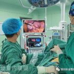
Minimally invasive treatment of intestinal fistula: the center has implemented a number of laparoscopic intestinal fistula operations, which can achieve the same effect as laparotomy in separating adhesion and exposing fistula, and have certain advantages in reducing postoperative pain and promoting patients’ early out of bed activities.
Thanks for reading, we hope you have enjoyed the content.
If you are looking for the endoscopy equipment, feel free to check our product page or contact us at our contact page.


Leave a Reply
Want to join the discussion?Feel free to contribute!